"Esophageal cancers are the eighth most common cancer and the sixth most common cause of cancer-related death."

Diagnosis


After conducting a physical exam and reviewing your health and family history, your oncologist will order various tests to diagnose and analyze your condition. An accurate diagnosis helps your medical team determine the best course of treatment. The most common tests for esophageal cancer include:

Imaging
  • Barium swallow, which can help to localize the level of obstruction.
  • Imaging tests, such as CT scan, PET-CT scan, and endoscopic ultrasound that will show a detailed image of esophagus, along with the depth involved, and also regarding local disease and other parts of the body.
Pathology
  • Endoscopic guided biopsy for histopathology, immunohistochemistry evaluation for confirmation of diagnosis will be done. If needed, molecular analysis including next genome sequencing can be advised.
  • Liquid biopsy
Staging of Esophageal Cancer

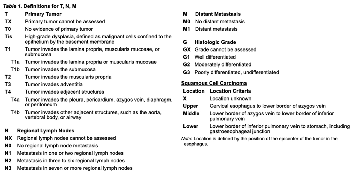
The staging system for Esophageal cancer is based on the American Joint committee on Cancer (AJCC) TNM system, 8th edition, which is based on tumor stage which relies on size, and invasion of nearby structures; nodal disease and distant metastases, which is based on tumor spread to other parts of the body. The staging system is the same for both SCC and AC.


The prognosis of the disease depends upon the stage at diagnosis

  • Endoscopic ultrasound to localize the extent of the disease confined to the esophagus and to the paraesophageal tissues
  • PET CT Scan to localize the nodal disease and for metastasis
  • Very rarely Laparoscopy in case of EGJ tumours


Treatment


Surgery

Surgery is a major component of treatment for locoregionalesophageal and esophagogastric junction (EGJ) cancers. Improvements in staging techniques, patient selection, post-surgical care, and surgical experience have led to a marked reduction in surgical morbidity and mortality in recent years. Additionally, randomized trials have shown that preoperative chemoradiation and perioperative chemotherapy have significantly improved survival in patients with resectable, locoregionally advanced esophageal and EGJ cancers.

What is unique in Surgery at APCC?

We perform Video-assisted thoracic surgery (VATS) and Robotic-assisted thoracic surgery ( RATS) for oesophageal cancers where the patient can go home in a week and lead a normal life.


The advantages of VATS and RATS


  • Less pain
  • Early recovery
  • Early resumption of routine activity
  • Less number of hospitals stay
  • Early adjuvant treatment
  • Less analgesic requirement
  • Less morbidity
  • Minimal trauma
  • Lesser tissue handling
  • More precise and cutting edge technology
  • Less mortality
Surgical Approaches

The type of esophageal resection depends upon the location of the tumour as well as the choice of conduit available. Two of the most common surgical approaches used are transthoracic and trans Hiatal esophagectomy, given below.

Transthoracic Esophagectomy

Ivor Lewis esophagectomy (right thoracotomy and laparotomy) and McKeownesophagectomy (right thoracotomy followed by laparotomy and cervical anastomosis) are the two standard options for transthoracic esophagectomy. Ivor Lewis esophagectomy, the most frequently used procedure for transthoracic esophagectomy, uses laparotomy and right thoracotomy, with upper thoracic esophagogastric anastomosis at or above the azygos vein.


Mobilization of the stomach for use as a conduit is performed, with dissection of the celiac and left gastric lymph nodes, division of the left gastric artery, and preservation of the gastroepiploic and right gastric arteries. This approach may be used for distal thoracic lesions, but the proximal esophageal margin will be inadequate for tumours in the middle esophagus. McKeownesophagectomy, with an anastomosis in the cervical region, is similar in conduct, but with the advantage of being applicable for tumours in the upper, middle, and lower thoracic esophagus.

TranshiatalEsophagectomy

Transhiatalesophagectomy (laparotomy and cervical anastomosis) is performed using abdominal and left cervical incisions. Mobilization of the stomach for the conduit is as done in Ivor Lewis procedure. This procedure is completed through the abdominal incision, and the gastric conduit is drawn through the posterior mediastinum and exteriorized in the cervical incision for the anastomosis. It can be used for any thoracic lesions, however, large middle esophageal tumours adjacent to the trachea will be technically challenging.

Transthoracic or ThoracoabdominalEsophagectomy

Left transthoracic or thoracoabdominalesophagectomy uses a contiguous abdominal and left thoracic incision through the eighth intercostal space. Mobilization of the stomach is as described, and esophagectomy is done through a left thoracotomy. The anastomosis is performed in the left chest, usually just superior to the inferior pulmonary vein, although it can be performed higher. It is usually used for lesions in the distal esophagus, particularly bulky tumours.

Minimally invasive Esophagectomy (MIE)

MIE includes minimally invasive Ivor Lewis esophagectomy and minimally invasive McKeownesophagectomy, limited laparotomy/laparoscopy, and cervical anastomosis. MIE strategies may be associated with decreased postoperative mortality and shorter recovery times.

Endoscopic therapies

Endoscopic therapies including Endoscopic Resection (ER-usually EMR, endoscopic mucosal resection and ESD, endoscopic submucosal dissection) and endoscopic ablation (cryoablation or RFA) have been used as alternatives to surgery for the treatment of early-stage esophageal and EGJ cancers. They have been associated with very less treatment-related morbidity compared to surgical resection. Early-stage disease like pTis, pT1a, select superficial pT1b without lymphovascular invasion can be effectively treated with ER and/or ablation.

Surveillance

Endoscopic surveillance following treatment of esophageal and EGJ cancers requires careful attention to detail for mucosal surface changes and multiple biopsies of any visualized abnormalities. It includes a search for the presence of Barrett esophagus and four-quadrant biopsies to detect residual or recurrent dysplasia. This option has to be chosen after careful consideration and the informed decision between you and your oncologist.

Radiation Therapy

Historically, radiotherapy alone was considered to treat esophageal cancer patients with unfavorable features, who are not medically fit for surgery. Overall, the 5-year survival rate for patients treated with radiotherapy alone was about 0-10%. Also, conventional means of radiotherapy is associated with higher organs at risk doses thereby causing more acute and late toxicities. The role of Intensity-modulated radiotherapy (IMRT), was investigated in the early part of the 21st century, in the treatment of esophageal cancer. Retrospective studies comparing 3D conformal radiotherapy (3DCRT) and IMRT have shown superior dose conformity and homogeneity as well as reduction of RT doses to organs at risk, especially the lungs and heart with IMRT. Also, IMRT along with concurrent chemotherapy in the definitive treatment of esophageal cancer yielded good results with acceptable toxicity profiles. A study in postoperative IMRT with concurrent chemotherapy for node-positive esophageal SCC showed the regimen to be safe and effective with one year overall and progression-free survival (OS and PFS) to be 91.2% and 80.4% respectively. Trials are ongoing and overall results have been so far exciting with concurrent chemoradiation in these tumours.

Proton Beam Therapy

Proton beam therapy (PBT), which is a superior form of radiotherapy, offers further sparing of normal tissues thereby minimizing doses to the organs at risk. Proton beam therapy, have a minimal exit dose beyond the target volume, which limits exposure to adjacent organs to radiation. A phase IIB trial comparing IMRT and PBT showed that PBT reduced the risk and severity of adverse events while maintaining similar rates of 3-year PFS (50.8% for IMRT and 51.2% for PBT) and 3-year OS (44.5% for both). PBT was also associated with lower rates of postoperative complications including pulmonary, cardiac, GI and wound complications, as well as reduced length of hospital stays. An ongoing phase III study comparing PBT to photon therapy is currently ongoing.


Proton beam therapy evolved, starting from the conventional passive scatter technology, to the modern image-guided pencil beam scanning technique, which improves the doses to the target volumes and minimizing doses to the organs at risk. Apollo proton cancer centre, is equipped with the latest image-guided Intensity-modulated proton therapy (pencil beam scanning technology with a spot size of 3mm, being the finest), along with superior Monte Carlo algorithm, which in turn will translate into better dose conformity to the target volumes and adjacent organs at risk.

What to expect if I choose proton beam therapy at APCC?

Your oncologist will first evaluate with a physical examination, historic reports and further investigations, and based on your stage of the disease, treatment options will be tailored along with a panel of experts (radiation oncology, medical oncology, surgical oncology). If you are planned for definitive or neoadjuvant or adjuvant radiation therapy using proton beam therapy, the following can be expected before and during treatment.


  • Informed consent will be taken explaining to you the benefits and side effects of radiation therapy/proton beam therapy.
  • Immobilization devices like Vacloc or mask will be done to minimize movements during precise radiation delivery.
  • A planning CT scan will be done with or without contrast, to delineate target volumes.
  • A 4D CT scan or a volumetric MRI may be acquired based on oncologist discretion, to assess your movement as well as the extent of local disease.
  • Further planning will include a minimum of one week with our group of expert medical physicists for precise dose delivery.
  • Your treatment will be initiated with a door to door time of approximately one hour.
  • Weekly reviews with your oncologist, with quality assurance CT, scans being done at the discretion of the oncologist as and when required.
Chemotherapy

Chemotherapy alone is usually not considered in the treatment of esophageal cancers except in a palliative or second-line setting. Chemotherapy is usually considered along with surgery in neoadjuvant or during perioperative setting, or along with radiotherapy, in preoperative, definitive or as adjuvant therapy. Several landmark trials like the CROSS protocol have shown a OS benefit in esophageal cancers with preoperative chemoradiotherapy followed by surgery. Your oncologist will explain the various drugs available to be used during treatment based on your treatment plan.

Palliative stenting

In patients who are medically unfit and want to have immediate relief of dysphagia, stenting can be attempted to have immediate relief and then proceed with second-line management. Stenting is usually palliative and is considered in advanced and metastatic diseases to improve quality of life.

Supportive care

People with esophageal cancer often experience both symptoms of cancer as well as side effects of treatment. Supportive care is essential in the treatment process. Our multi-faceted team is focused on ensuring patient comfort and better quality of life.

Follow up

Patients completing treatment for esophageal cancers will be advised regular follow ups, once every three months for the first two to three years, six-monthly during fourth and fifth years, and yearly thereafter. APCC is committed to excellent follow-ups, with state of art digital PET-CT scan and multidisciplinary board discussions. We have robust teleconsultation and video consultation for ensuring timely follow up in these difficult Covid times.

Winning Over Cancer

Mr Mohammed Jamal Uddin from Bangladesh shares his experience about undergoing Proton treatment for his lung tumour at Apollo Proton Cancer Centre.

Watch him speak about his journey.

FOR APPOINTMENT

Mr. Praboth Palit from Mumbai shares an emotional journey of his treatment at Apollo Proton Cancer Centre. Despite facing a lot of challenges in the beginning, Praboth never lost his hope which is highly commendable.

He thanked Mr. John Chandy , Dr. Srinivas Chilukuri, and the entire team for their support and guidance.

FOR APPOINTMENT