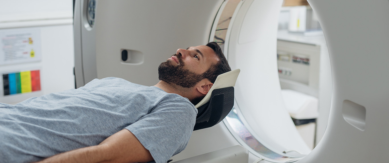
Accurately Diagnosing Cancer: The First Step in Treatment
The treatment process at APCC begins with best-in-class diagnostics. A confluence of cutting-edge technology, high skill and great expertise give us a definitive edge in this vital area of cancer care. Our focus on diagnostics is imperative for our mission of better cancer care. As knowledge about the genetic and molecular profile of each cancer becomes increasingly incremental, having an in-depth understanding of mutational load and profile becomes critical.
In healthcare, knowledge is empowerment. Identifying the exact problem is the beginning of the process of optimal treatment. Identifying the ailment provides clarity of purpose and direction, which are mission-critical in the process of cure. Therefore, accurate diagnostics are the bedrock of world-class clinical outcomes.
Experience the latest in high-end diagnostics at APCC. From advanced imaging technology to cutting-edge lab tests, our comprehensive cancer diagnostic services provide accurate & timely results for a range of medical conditions.
Common diagnostic tests include:
- Laboratory Tests: To examine the presence of certain substances in your blood or urine.
- Imaging Tests: Includes CT scan, X-ray, ultrasound, MRI, PET scan and Nuclear medicine scan. These tests enable radiologists to examine suspicious areas by producing highly detailed pictures of the inside of your body.
- Endoscopy: To view areas inside the body via a tiny camera on the end of a flexible plastic tube that is inserted into body cavities and organs.
- Genetic Tests: Use a patient’s blood sample to look for genetic mutations that may lead to an increased risk for some cancers. After a medical and family history review, a genetic counsellor will discuss whether genetic testing is right for you.
Imaging Tests
3 Telsa Magnetic Resonance Imaging (MRI)
MRI stands for Magnetic Resonance Imaging. It is a diagnostic imaging technique that uses a magnetic field and radio waves to produce detailed images of your body's internal organs and tissues. The unit of measurement used to quantify the strength of a magnetic field in an MRI machine is called a Tesla. Most MRI scanners come in the 1.5 Tesla range. At APCC, we have the revolutionary 3 Tesla MRI. Operating at twice the power, it provides a greater signal-to-noise ratio, which is a major determinant in generating the highest quality image.
The higher resolution images produced by the 3 Tesla MRI are beneficial when diagnosing pathological conditions involving the brain, spine, and musculoskeletal system. The resolution and clarity also allow radiologists to identify smaller lesions and anatomical structures that cannot be seen with less powerful machines.
Advanced Mitral Regurgitation (MR) Techniques:
1. Diffusion-Weighted Imaging (DWI)
DWI is a short technique used to detect acute ischemia in the brain parenchyma and is an integral part of neuroimaging. DWI data processing helps in obtaining an Apparent Diffusion Coefficient (ADC) maps. The ADC values have an inverse relation to the cellularity and grade of glioma. ADC values are also low in tumours such as primitive neuroectodermal tumours and lymphomas. DWI can be diagnostic in non-neoplastic conditions such as epidermoids and cholesteatomas as well as pyogenic abscesses. DWI can be used for preoperative grading of brain tumours, predicting cellularity, directing the site of biopsy, differentiating between recurrent disease and radiation effects, and in the assessment of treatment response. In the postoperative imaging, DWI can delineate residual tumour tissue with high cellularity as well as the ischemia resulting from the intervention.
2. Diffusion Tensor Imaging (DTI)
DTI is an advancement of Diffusion-Weighted Imaging and provides information of the tracts in the brain. It can provide important preoperative information regarding contiguous white matter fibers. This information provided by DTI enables the surgeon to decide the safest surgical approach, particularly in tumors involving the brainstem and those in proximity to the internal capsules. A fusion of data obtained on fMRI and DTI is advantageous for pre-operative planning of tumour excision in proximity to the eloquent cortex. DTI of the spinal cord is also performed to assess the integrity of the spinal tracts.
3. MR Spectroscopy
MR spectroscopy relies on the MR phenomena of chemical shift and spin-spin coupling effects to provide important biochemical information. The main metabolites of interest in brain tumours include:
- N-acetyl aspartate (NAA), a marker of neuronal integrity;
- Choline (Cho), a marker of cellular membrane turnover;
- Creatine (Cr), a marker of bioenergy stores;
- Lactate (Lac), a product of anaerobic glycolysis;
- Lipids (Lip), by-products of necrosis;
- Glutamate-glutamine, and Gamma-aminobutyric acid (Glx and GABA), neurotransmitters;
- Myoinositol (mI), a glial cell marker.
Creatine is used as an internal reference marker for cellular metabolism. And, if present, lactate may be interpreted as a marker for hypoxia, while the presence of mobile lipids within a tumour indicates necrosis. Both represent additional characteristic features of malignancy. The presence of certain metabolites may be detected in particular tumour subtypes, such as alanine in meningioma, taurine in medulloblastomas or amino acids in pyogenic abscesses. In tuberculomas, an important differential diagnosis of ring-enhancing lesions in tropical countries, the lipid-lactate peak is tall and reflects the caseation within. Choline peak may be slightly increased in chronic lesions due to the cellularity caused by aggregated macrophages.
In addition to helping in confirming the diagnosis, MR spectroscopy can be used in the grading of lesions. Higher choline and lipid-lactate levels indicate a higher grade and a preserved spectrum; a slight increase in choline and absent/minimal lipid-lactate levels indicate a lower grade. In surveillance imaging of high-grade gliomas, the height of the choline peak is expected to decrease as a response to the treatment. In recurrent glial tumours, choline increases, while as radiation effect, choline can be expected to be low, though there is a considerable overlap of these features. The interpretation then depends on DSC and 3D ASL perfusion studies, in addition to the morphology of the abnormality on the conventional images.
4. Perfusion Weighted Imaging (PWI)
PWI is an MR technique, which detects variations in the hemodynamics of the blood flowing in the brain. PWI can be performed with or without injecting an exogenous contrast. Perfusion techniques involving the injection of contrast include Dynamic Susceptibility Contrast (DSC), and Dynamic Contrast-Enhanced (DCE).
In the commonly used DSC perfusion technique, a contrast agent is injected rapidly as a bolus into the venous system. To assess the passage of the agent across the blood-brain barrier, echoplanar imaging is used. BV (Blood Volume), BF (Blood Flow), MTT (Mean Transit Time), and TTP (Time To Peak) maps are obtained. rCBV ratios have a direct relation to the grade of glial tumours, even in non-enhancing high-grade tumours and can indicate the site of biopsy by outlining the aggressive areas within a heterogeneous tumour bed. Effects of radio and chemotherapy treatment, including pseudoprogression or necrosis, demonstrate lower CBV values. Steroids and anti-angiogenic drugs such as Bevacizumab may lower the rCBV ratios, despite lack of response. The knowledge of these eventualities helps in better interpretation.
5. Arterial Spin Labelling (ASL)
ASL is a novel technique. It can be used as an alternative or as an addition to DSC in the majority of cases, especially on our new generation 3 Tesla Magnetic Resonance (MR) scanner. Three-dimensional pseudo-continuous ASL (3D pcASL) is the technique found to be most sturdy by experts. The technique uses an endogenous tracer, namely the water in the flowing arterial blood, and hence does not require the use of contrast. This is beneficial for patients with compromised renal function, repetitive follow-ups of post-chemotherapy patients, and in children, for whom fast injection for DSC is difficult. ASL can measure absolute values and is not affected by permeability, along with being less sensitive to variations caused by hemorrhage. The utilization of 3D ASL in routine imaging of brain tumours, with accurate post labelling delay, which varies according to the age of the patient is encouraged.
ASL has been very useful in brain tumour assessment, in both pre and post-operative setting – to grade the tumours, to localize the site of biopsy, to assess post-operative residual neoplastic activity, differentiate recurrence and post-treatment changes, and to assess the efficacy of newer treatment modalities, like anti-angiogenic drugs.
6. Functional MRI (fMRI)
fMRI is a technique that measures alterations in the oxygenated and deoxygenated blood during neuronal activation while performing a specific task, and at rest. When a task is performed, there is increased blood flow in the region of the neuronal activity, which results in a decrease in the deoxygenated hemoglobin, as oxygen brought in is in excess. Deoxyhaemoglobin has paramagnetic properties, and the resultant distortion of the magnetic field can be measured as a small signal. This technique is called Blood Oxygenation Level-Dependent (BOLD) contrast. The fMRI data obtained is then registered onto high-resolution MR anatomical images acquired without moving the patient. This data, along with DTI, can be transferred to the neuro-navigating system in the operative suite.
The role of functional MRI in neuroimaging includes the mapping of the eloquent areas and establishing their relationship to the margins of tumours or other intracranial lesions, and assessing the side of cerebral dominance.
Computed Tomography (CT) Scan
A CT scan uses an X-ray machine to create a 3-dimensional picture of the inside of the body. A CT scan uses a pencil-thin beam to create a series of pictures taken from different angles. A computer then combines these images into a detailed view that shows any abnormalities or tumours.
CT scans show a slice or cross-section of the body. The image generated shows bones, organs, and soft tissues in greater clarity than a traditional X-ray. CT scans can show a tumour’s shape, size, and location. They can even show the blood vessels that feed the tumour.
Nuclear Medicine
Nuclear medicine is a medical specialty that involves the use of radioactive materials or radiopharmaceuticals for the diagnosis and treatment of diseases. Nuclear medicine scans provide information on both the anatomy and the physiology of the body. They also help determine the presence of disease based on the function of the organ, tissue or bone.
With the aid of radioactive material, our team at APCC can assess organ function, evaluate for the presence of disease, and effectively treat certain conditions. For diagnostic purposes, the radiopharmaceuticals emit photon radiation that is detected by special cameras to produce images which are read by the physicians. For therapeutic purposes, the radiopharmaceuticals emit particle radiation which can cause biological damage, injuring, or destroying cancer cells. At APCC, the Department of Nuclear Medicine is an integrated and aligned team of professionals with different niche expertise, all with the common goal to lead clinical, educational, and research endeavors in the effort of providing optimal cancer care.
Positron Emission Tomography And Computed Tomography (PET-CT) Scans
PET-CT is a well-established diagnostic tool for cancer where radiolabelled PET radiotracers, most commonly 18F-Fluoro-2 deoxyglucose (18F-FDG), a radioactive sugar molecule is injected into the patient. Cancer cells metabolize sugar at higher rates than normal cells and a PET-CT scan helps the physicians to understand if the tumour within the body is cancerous or not. The PET-CT study provides information regarding the stage of the disease, so that appropriate management such as surgery or chemotherapy or radiation therapy can be advised, as deemed appropriate.
Apollo Proton Cancer Centre is equipped with the new age 64 slice Siemens Biograph Vision, the first Digital PET-CT scanner in South East Asia. The FDG-PET-CT in the Apollo Proton Cancer Centre can also help in the early detection of tumour recurrence. PET-CT scans are now increasingly being used for monitoring treatment response across various malignancies, which will be able to monitor the effects of therapy and change the same in case the patient does not respond to it.
Advantages of PET-CT
- Early detection of disease before the appearance of structural changes.
- Accurate staging and restaging of cancers.
- Simultaneous contrast-enhanced 64 slices whole-body CT with metabolic information.
- Quantification of tumour metabolism before and after treatment.
- Metabolic grading of tumours and prognostication.
- Safe for use in patients with chronic kidney disease, metallic implants, pacemakers, etc.
Patient-Centric Features
- Wide gantry bore size (78 cms): accommodates larger patients.
- Short gantry tunnel (136 cms): improves patient comfort and facilitates patient positioning.
- Zero deflection of the bed between PET and CT fields of view: the smooth motion of the patient throughout the procedure.
- Higher weight-bearing capacity: Upto 227-kgs
Special Features: The most advanced crystal in India with digital detectors with the ability to provide;
- Highest sensitivity: Reduces scan time and injected dose, improves patient throughput, and reduced radiation exposure in patients.
- Best in the industry resolution: Can detect small lesions and impact early evaluation of treatment response.
- Fastest time of flight technology (214 ps): Improves contrast and signal to noise ratio.
- Flowmotion and multi-parametric PET imaging: enables absolute quantification compared to SUV alone. Enables whole-body dynamic images.
- Fast reproduction from original data.
- 4DCT for respiratory gating: For accurate radiation therapy planning.
- Care does 4D: Automatic CT settings to minimize radiation exposure.
Dual-Energy X-Ray Absorption (DEXA) Scan
A DEXA scan is a test that measures bone mineral density. It helps in evaluating bone health and determining the likelihood of osteoporosis or bone fractures. This scan may also help in detecting if cancer has metastasized or spread to the bones.
For a patient diagnosed with cancer, doctors may perform a baseline DEXA scan before and during treatment to monitor their bone health. Based on the results, measures will be taken to help prevent bone loss. The quick and painless scan uses low levels of X-rays to measure bone mineral density. The patient is made to lie on a table for 15-20 minutes as their entire skeleton and/or specific points on the body are scanned. Once the scan is complete, the results are composed of two different scores:
T-Score: The difference between the patient’s bone density and that of an average healthy person. This score is used to determine the patient’s risk of breaking a bone.
Z-Score: The amount of bone the patient has compared to other people of the same age, race and gender. A score that is too high or too low may require more testing.
Digital Radiography
Digital Radiography is an innovative departure from the conventional method of using film to record images. Here, the images are captured and stored in digital formats that can be stored, manipulated and shared very easily. It has been proven to be vastly superior to the standard film-based radiography. Digital Radiography typically reduces radiation exposure by 75% or more and is emblematic of a higher quality of care. Digital X-ray systems help us control the exposure of each image in real-time so that images can be made darker or lighter on demand. It also makes it much easier to enlarge images, enhance colours, and superimpose textures.
Mammography
Mammography is an imaging modality that checks for breast cancer in women. The images that it produces are called mammograms. These images may show small tumours that cannot be felt, along with other irregularities in the breast. A mammogram could be either to screen for breast cancer or to diagnose it. Mammography is a rapidly evolving field. Digital Mammography, which records the images on a computer rather than on film, produces sharper, high contrast images that can help highlight important details. Another advance is breast chemosynthesis – a technique that generates 3D images of the breast.
Sonography
Sonography, using ultrasound, is an important detection methodology in cancer diagnosis. The ultrasound machine bounces sound waves off organs and tissues. These waves create echoes that are converted into real-time pictures that show organ structure and movement. Sonograms can even track blood flow through blood vessels. Ultrasound is highly beneficial in getting pictures of soft tissue diseases that don’t show up well on X-rays. At APCC, we use sonography to investigate and differentiate fluid-filled cysts from solid tumours because of their distinct echo patterns.

Winning over Cancer with Apollo Proton Cancer Centre
A breakthrough in Cancer Care! The global growing cancer burden tells an ominous tale. To counter this growing threat, Apollo Proton Cancer Centre provides a complete and comprehensive solution. As cancer care has become one of the fastest-growing healthcare imperatives across the world, we believe it is critical to redefine our purpose, to reboot our commitment on the single-minded focus - to battle cancer, to conquer cancer! APCC stands as a ray of hope for millions, infusing them with the courage to stand and stare cancer down.

Copyright © 2023 Apollo Proton Cancer Centre. All Rights Reserved


